DNA (deoxyribonucleic acid), the giant
molecule that carries genetic information in living things, is made up of just a few
chemical building blocks that bond together in very particular ways.
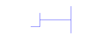
A typical molecule of DNA consists of two
strands that are linked together. A segment can be visualized as a ladder-like structure:
(Actually DNA looks like a twisted ladder or
a spiral staircase, a shape commonly called a helix or, since there are two strands in the
structure, a double helix. Here we are focusing just on the structure of the ladder.)
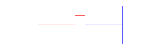
In our flat unrealistic model picture we are visualizing the DNA as made of two strands, a left and a right,
which are linked in the middle. In reality, since the ladder is twisted, there really is
no "right" or "left," but we use that terminology here since it
applies to the model graphs. The square shapes and diamonds you see in the middles of the
"rungs" are the links, which are meant to represent the different chemical bonds between the two strands
of DNA.
Each of the two strands of DNA consists of a sequence of nitrogenous bases, and any one of
four such bases can appear on a strand. (The DNA also contains other molecular building
blocks like sugars and phosphate groups, but the nitrogenous bases are the genetically
important part.) The four bases and our geometric representation of each are as follows:
Base
Representation
adenine
thymine
cytosine
guanine
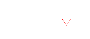
These four bases are usually referred to by
their leading initials: C (cytosine), G (guanine), A (adenine) and T (thymine). The shapes
used here were selected intentionally. Note what happens when a C appears on the left
strand . . .
. . . and a G in the corresponding spot on
the right strand . . .
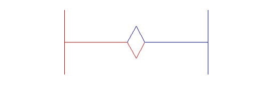
The two bases hook up nicely!
The solid bond the two bases form is
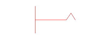
indicated by the closed solid in the middle. Similarly with A and T:
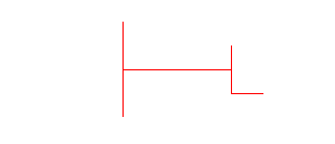
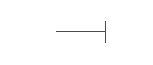
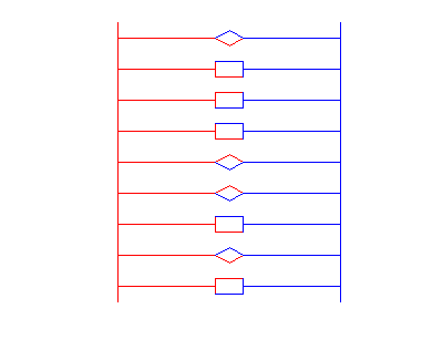
In the construction of DNA, C can only be
matched with G and A only with T. All four can be part of a strand. Pictorially you can
see that here when the bases just don't match up . . .
. . . or simply do not form a closed bond .
. .
Thus once you specify
the sequence of bases on one strand, the sequence on the other is determined.
For example, if the left strand is
then the right strand
must be
Graphically, we can
represent the resulting piece of DNA as
You can modify the vectors L and R above as
much as you like to help reinforce these concepts. Make L as long as you wish and enter a
sequence of As, Gs, Cs and Ts. Then fill in the vector R of the same length with the
correct right strand sequence. Inspect the resulting graph to see if everything bonded
correctly.
FREE
Software (pDRAW32) to draw DNA Analysis Chartshttp://www.acaclone.com/ pDRAW32 lets you enter a DNA name and
coordinates for genetic elements, such as genes, to be plotted on your DNA
plots. pDRAW32 lets you "clone" fragments of DNA generated by
virtual digestion with restriction enzymes and optionally blunted at one or both
ends. Up to 3 fragments may be cloned at a time (can you replicate that in the
lab?). Each fragment may be inverted relative to its original orientation.
Genetic elements contained in the cloned fragments are transferred to the cloned
DNA. (...and much more!)
Procedure to ligate fragments of genomic DNA from spinach into a
vector plasmid; this recombinant DNA is then used to transform Escherichia
coli cells.
NEW! FREE
Software (pDRAW32) to draw DNA Analysis Charts http://www.acaclone.com/
pDraw32 DOWNLOAD software - pDRAW32 lets you enter a DNA name and
coordinates for genetic elements, such as genes, to be plotted on your DNA
plots. pDRAW32 lets you "clone" fragments of DNA generated by
virtual digestion with restriction enzymes and optionally blunted at one or
both ends. Up to 3 fragments may be cloned at a time (can you replicate that
in the lab?). Each fragment may be inverted relative to its original
orientation. Genetic elements contained in the cloned fragments are
transferred to the cloned DNA. (...and much more!)
Procedure to ligate fragments
of genomic DNA from spinach into a vector plasmid; this recombinant DNA is
then used to transform Escherichia coli cells.